Mostly the alternate pathway of complement activation is involved.
(Evidence – decreased serum C3 level and normal C1,C2 and C4).
The deposits of C3 (granular pattern) is seen in glomeruli.

C3NeF (Circulating anti complementary nephritic factor) is an Autoantibody (Ig) – binds to C3 convertase and stabilise it leading to its persistent activation.
EPITHELIAL INJURY
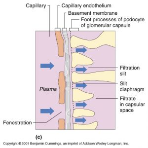
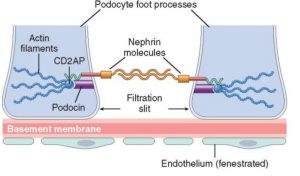
Visceral epithelial cells (PODOCYTE) bears FOOT PROCESS which serves the function of formation of filtration slit and hence prevent protein filtration in glomerulus.


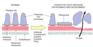
Epithelial cells may get injured by the antibodies, toxins and cytokines.
These cells have low regenerating capacity,
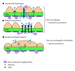
The injury is reflected by changes in the epithelial cells like detachment of foot process (because of loss of adhesion with basement membrane), impaired slit diaphragm etc.
Finally this injury leads to loss of function of epithelial cell and leakage of proteins occurs causing PROTEINURIA.
The clinical entities which show this pattern of injury is MINIMAL CHANGE DISEASE, DIABETIC NEPHROPATHY.
PATHOGENESIS OF MINIMAL CHANGE DISEASE


