
Imp features –
Largest salivary gland.
Situated between ESR– external acoustic meatus, ramus of mandible & sternocleidomastoid.
Capsule –
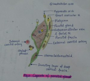
Formed by investing layer of deep cervical fascia, which splits to enclose the gland.
Capsule – Superficial and Deep Lamina
- Thick Thin
- Adherent to gland.
- Attached to styloid process, Zygomatic arch, tympanic plate.
External features –
- Resembles an inverted 3 sided pyramid.
Has 4 surfaces – superior, superficial, anteromedial and posteromedial.
Has 3 borders – anterior, posterior, medial.
Structures within the gland –
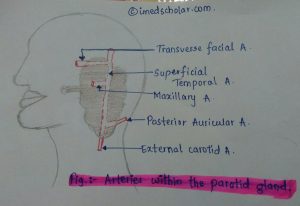
Arteries –
External carotid A. enters the gland and maxillary artery leaves the gland.
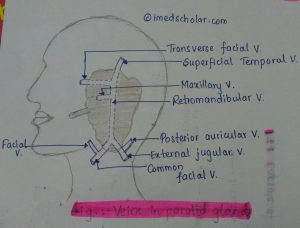
Veins –
Within the gland-
Superficial temporal vein.
Retromandibular vein.
Maxillary vein
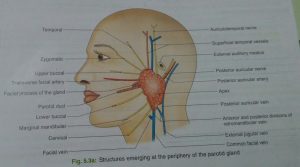
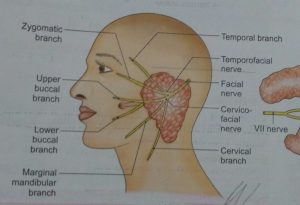
The Facial Nerve–
The Facial Nerve gives its terminal branches within the gland.
It divides into 2 branches
1.Temporofacial– Temporal
– Zygomatic
2.Cervicofacial – buccal
– marginal
– mandibular
– cervical
These branches radiate as goose foot from the anterior border of the gland.
Mnemonic –
To Zanzibar by motor car
T – Temporal
Z- Zygomatic
B – buccal
M – mandibular & marginal
C – Cervical
Parotid duct –
Also called Stenson’s duct
5 cm long
Opens into the vestibule of mouth opposite to the crown of upper 2nd molar tooth.
Blood supply –
Arterial supply – external carotid artery and it’s branches.
Venous drainage- veins drain into external & internal jugular veins.
Lymphatic drainage–
Lymph – Parotid nodes –upper deep cervical nodes
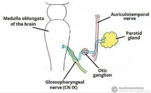
Nerve Supply

Mnemonic –
IT has Lesser Options Anywhere.
I – inferior salivatory nucleus
T – Tympanic branch of 9th nerve
Lesser –lesser petrosal nerve
O – otic ganglion
A – auriculotemporal nerve.
Interesting fact –
The facial nerve lies in the parotid gland and divides into branches but does not innervate the parotid gland!

Important Clinical anatomy-
After removal of parotid gland, there is regeneration of secretomotor fibres in auriculotemporal nerve , causing stimulation of salivary glands & hyperemia, producing redness & swelling – Frey’s Syndrome.
The parotid gland is removed in 2 parts to preserve facial nerve.