Let’s first understand what it means.
ACUTE– transient in nature (rarely leads to renal failure)
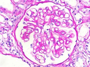
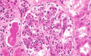
PROLIFERATIVE– proliferation of endothelial and mesangial cells
POST– 1-4 weeks after
INFECTIOUS– Occurring because of infections like staphy, strept.
GLOMERULONEPHRITIS– Inflammation of glomerulus.
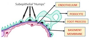
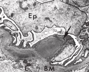
The basic mechanism which underlies this disease is SUBEPITHELIAL immune complex deposition.
So, just let’s go to basics. What an immune complex is formed of?
Obviously, an antigen and its specific antibody reacting to form it.
Here, the antigen may be endogenous (Ex. SLE) or exogenous (via some infecting organism like strept, staphy, HBV, HCV, HIV, pneumococcus).
The most common Ag involved is of streptococcus. On this basis the APGN is divided into 2 categories- POST STREPTOCOCCAL GLOMERULONEPHRITIS (PSGN) and NON STREPTOCOCCAL GLOMERULONEPHRITIS.
So basically we will focus on PSGN.
It is caused by group A beta haemolytic nephritogenic streptococcus. The most important Ag involved in PSGN is SpeB (streptococcus pyogenic exotoxin B).
Other Ag involved are- antistreptolysin O, antiDNAase B etc.
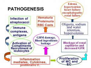
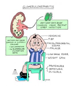
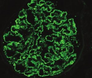
What happens is that following the infection of streptococcus, antibodies (IgG) are formed in the body within 1-4 weeks which forms immune complex and gets deposited in the glomeruli and causes complement activation leading to C3 deposits as well as elicit inflammatory response leading to leucocytic infiltration.
Note that the antibody formed in staphy is of IgA type
The disease is most common in children (6-10yrs).
The typical history is that a child developed sore throat and 1-4 weeks later experienced malaise, fever, nausea, oliguria, haematuria and mild proteinuria. It has a good prognosis with 95% recovery. Only 1% patient develops RPGN.
Prolonged, persistent and heavy proteinuria- UNFAVOURABLE DIAGNOSIS
-Mansi Nahar, GMC Nagpur

