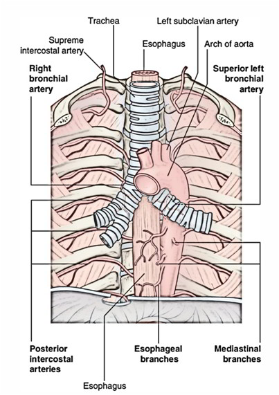
The ascending aorta extends itself as the arch of aorta at sternal angle and it further extends itself as descending thoracic aorta at sternal angle. So, the beginning and the ending is at the level of sternal angle. It is situated in the superior mediastinum behind the lower half of manubrium sterni.
- Begins as a continuation of the ascending aorta behind the right second chondosternal joint.
- Passes up, back and left in front of the bifurcation of the trachea.
- Finally turns backward and downwards behind the left bronchus and continues as descending aorta at lower border of T4 vertebra.
ANTERIOR AND TO THE LEFT
- Left lung and pleura.
- Left phrenic nerve.
- Left vagus nerve.
- Left cardiac nerves.
- Left superior intercostal vein.
Friends(phrenic) played cards(cardiac) at the long(lung) coast(intercostal) in vagus.
INFERIOR
- Left bronchus.
- Ligamentum arteriosum along with superficial cardiac plexus.
- Left recurrent laryngeal nerve.
Bran (bronchus) watched La Liga (ligamentum) with Larry (laryngeal nerve).
SUPERIOR
- Brachiocephalic trunk.
- Left common carotid artery.
- Left subclavian artery.
- Left brachiocephalic vein.
- Remains of thymus.
Brick from the trunk (brachiocephalic trunk) of the brick van (brachiocephalic vein) fell on the thigh(thymus) of the common (common carotid) man in the subway (subclavian artery).
- AORTIC KNUCKLE
In X ray chest (PA view), the shadow of arch of aorta appears as small bulb like projection in the upper end of the left margin of the cardiac shadow termed aortic knuckle. The aortic knuckle may become notable in old age because of undue folding of the arch caused by atherosclerosis.
- COARCTATION OF AORTA
Congenital narrowing of the aorta just proximal or distal to the entrance of the ductus arteriosus.
- PATENT DUCTUS ARTERIOSUS
In foetal life, pulmonary trunk is connected to the arch of the aorta (just distal to the origin of left subclavian artery) by short wide channel termed ductus arteriosus. Non- obliterated ductus arteriosus is termed as patent ductus arteriosus.
- ANEURYSM OF THE ARCH OF AORTA
It is the localised dilatation of the arch and causes compression of neighbouring structures in the superior mediastinum producing mediastinal syndrome. The characteristic mediastinal sign in this condition is a tracheal-tuga that is a feeling of tugging sensation in the suprasternal notch.

