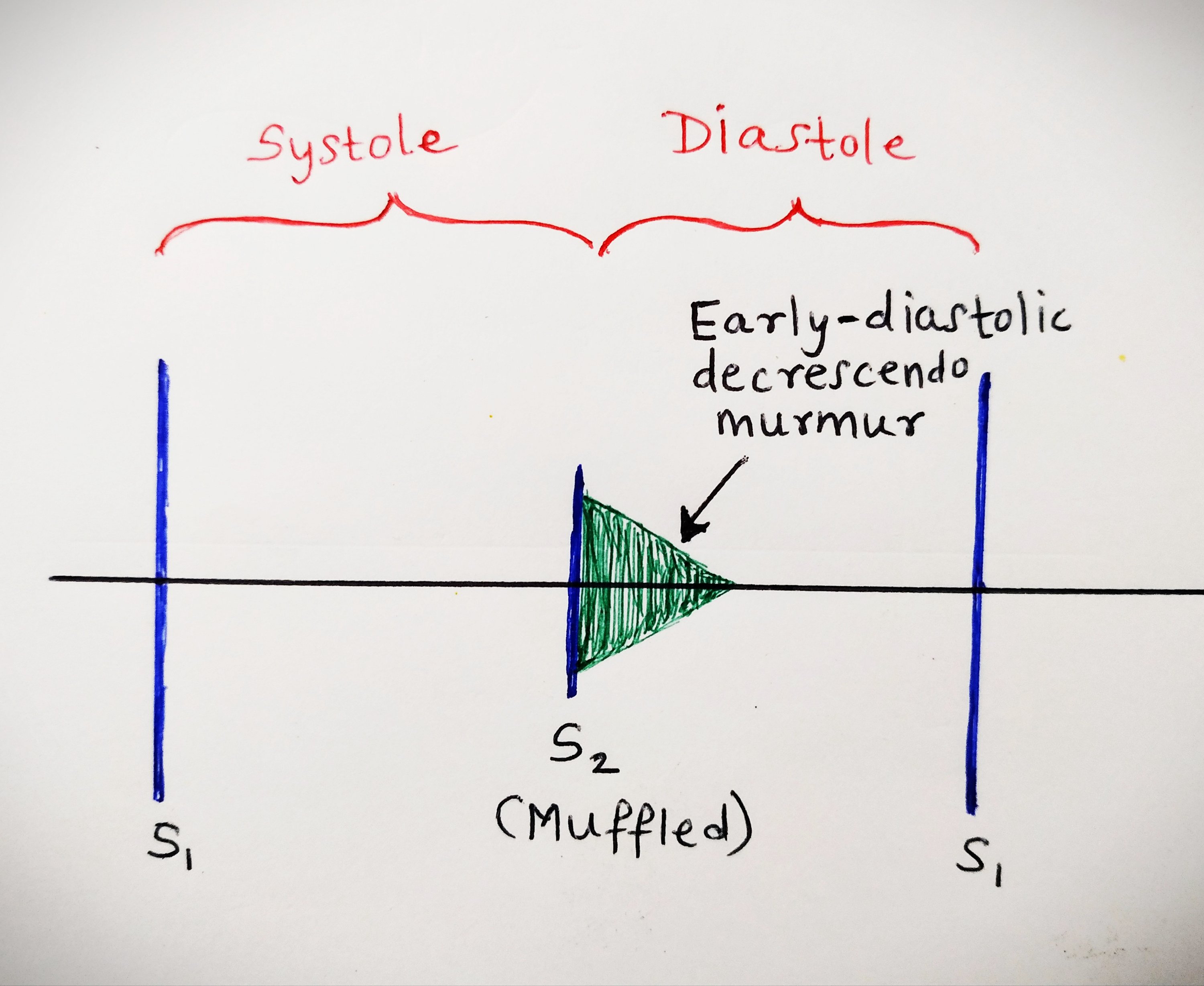
In aortic regurgitation, the aortic valve is incompetent. Hence, some of the blood which is pumped into the aorta from the left ventricle during systole, falls back into the left ventricle during diastole.
The left ventricle has to now work harder to pump out the excess blood and this leads to LVH and eventually LVF.
Causes of AR:
1.Rheumatic
2.Traumatic
3.Bicuspid aortic valve
4.Syphilis
5.Aortic dissection
6.Dissecting aneurysm of aorta
Symptoms of AR:
1.Palpitations
2.Throbbing sensation throughout the body
3.Chest pain (because of pounding of the heart against the chest wall)
4.Exertional dyspnea
5.Features of LVF or CCF
Signs of AR:
Pulse: Water hammer pulse, peripheral pulses are nicely felt
BP: High systolic and low diastolic BP (wide pulse pressure)
Apex: Hyperdynamic and diffuse (Felt in more than 1 intercostal space, or area more than 25 paisa coin)
Apex is shifted downward and outward because of LVH.
S2: Muffled
S3: may be heard sometimes
Classical Murmur of AR: Soft blowing, early diastolic, decrescendo murmur heard best at the neoaortic area (Erb’s area) with the diaphragm of the stethoscope in sitting and leaning forward position. The murmur is radiated towards the apex.
Occasionally, the murmur may radiate upto apex and left axilla, and is known as Cole-Cecil murmur.
Other types of murmurs found in AR:
• Ejection systolic murmur of functional AS in the aortic area or neoaortic area.
•Austin Flint murmur: mid diastolic rumbling murmur of functional MS (the regurgitant jet of blood strikes the mitral valve and stenoses it)
Features of pulmonary hypertension may be present if it develops as a complication

Auscultatory findings in Aortic regurgitation
Peripheral signs of AR- very important. (They are produced because of wide pulse pressure)
1.Quincke’s sign– Visible capillary pulsation at nail bed or lips
2.Prominent digital artery pulsation
3.Corrigan’s pulse –Waterhammer pulse or high volume collapsing pulse
4.Corrigan’s sign-Dancing carotids in the neck
5.BP-high systolic and low diastolic (Wide pulse pressure)
6.Locomotor brachialis- tortuous and highly pulsatile brachial artery
7.De Musset’s sign-To and fro head nodding synchronous with carotid pulsation
8.Visible pulsation in suprasternal notch
9.Traube’s sign: Pistol shot sound produced on pressing the stethoscope over the femoral artery
10.Systolic murmur on compression of the femoral artery proximally.
11.Duroziez’s murmur: Diastolic murmur on compression of the femoral artery distally
12.Hill’s sign: Increase in the femoral artery systolic BP > 20 mm of Hg above the brachial artery systolic BP
13.Pulsations in the uvula: Muller’s sign
14.Pulsations in the liver: Rosenbach’s sign
15.Pulsations in the enlarged spleen: Gerhardt’s sign
16.Lighthouse sign: Alternate flushing and blanching of forehead
17.Landolfi’s sign: changes in pupil sign with each cardiac systole
18.Pulsations of retinal artery: Becker’s sign
Complications of AR:
1.Acute LVF
2.CCF
3.Arrythmias
4.Infective endocarditis
5.Angina (mostly nocturnal)
6.Heart block
Investigations:
1.Chest X-ray
•Cardiomegaly (due to LVH) called Cor bovinum
•Dilatation of aortic root
•Calcified aortic valve
2.ECG: signs of LVH
3.Echocardiography
4.Blood test to detect active rheumatic carditis: TLC, DLC, ASO titre and to detect syphilis (VDRL, Kahn test)
5.Cardiac catheterisation
6.Doppler study (most sensitive test to detect mild AR)
Treatment of AR:
1.CCF: Diuretics, digoxin, salt restriction, restriction of physical activity
2.Treatment of underlying cause like syphilis or Rheumatic heart disease.
3.Chronic AR with volume overload: Vasodilator (nifidepine, hydralazine,ACE inhibitors)
4.Aortic valve replacement
Questions:
Why is the murmur in AR early diastolic?
Because the pressure gradient between the aortic valve and left ventricle is the highest in early diastole, hence there is leakage of blood into the left ventricle in early diastole.
In mid diastole, the pressure in the aorta and left ventricle becomes equal.
Classification of severity of AR on the basis of Hill’s sign.
20-40 mm Hg: Mild AR
40-60 mm Hg: Moderate AR
>60 mm Hg: Severe AR
Other similar posts:
