- The process of synthesizing ATP from ADP & Pi coupled with electron transport chain is known as oxidative phosphorylation.
- For this oxidation to take place, many components are required. One of them is NADH, the reducing equivalent.
Transport of reducing Equivalents – the shuttle pathways.
- The inner mitochondrial membrane is impermeable to most charged particles. It lacks NADH transporter.
- Therefore, NADH produced in the cytosol cannot directly enter the mitochondria.
- However, two electrons of NADH are transported from cytosol into the matrix of mitochondria using shuttle pathways.
- Malate Aspartate Shuttle
- Glycerol Phosphate Shuttle.
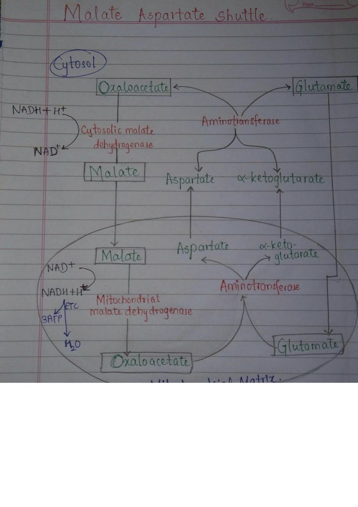
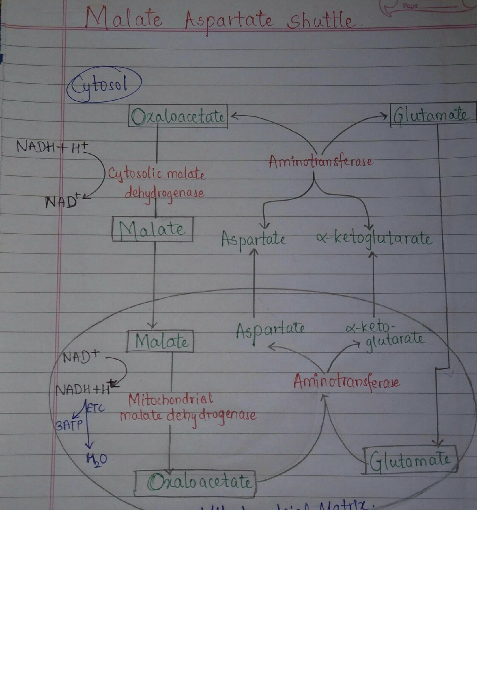
Malate Aspartate Shuttle
- In the cytosol, oxaloacetate accepts reducing equivalents (NADH) and becomes malate.
- Malate then enters mitochondria & gets oxidised by mitochondrial malate dehydrogenase . In this reaction, NADH & oxaloacetate are regenerated.
- NADH that enters the matrix gets oxidised via the electron transport chain & 3 ATP are produced.
- Now the oxaloacetate generated participates in transamination reaction with glutamate to produce aspartate &alpha- ketoglutarate.
- The aspartate & alpha – ketoglutarate then undergo transamination to give oxaloacetate & glutamate and the cycle continues.
- Thus, 3 ATP per mole of NADH is generated by Malate Aspartate shuttle.
- Another pathway, the Glycerol Phosphate Shuttle works to transport the reducing equivalent, FAD.
But, in this pathway, only 2 ATP molecules are generated.
- Liver & Heart utilize the malate- aspartate shuttle to produce ATP.
While other tissues use the Glycerol Phosphate Shuttle
- Regulation
The activity of malate-aspartate shuttle is modulated by arginine methylation of Malate dehydrogenase 1 (MDH1). Protein arginine N-methyltransferase CARM1 methylates and inhibits MDH1 by disrupting its dimerization, which represses malate-aspartate shuttle and inhibits mitochondrial respiration of pancreatic cancer cells.
Contributed by: Soumya Khot